Before After
 |
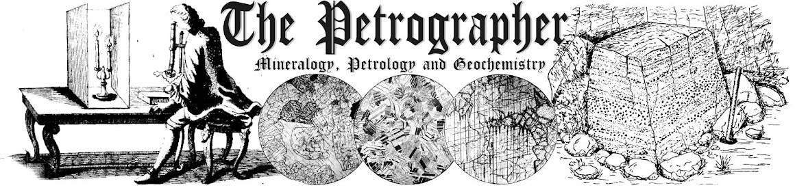
| Remains of amphibole in the ablation crater (center, right image) demonstrate relatively low birefringence color. The pictures were taken using different settings of camera and microscope, XPL. |
|
 |
| Closeup of the crater in the same spot. Reflected light. |
Imagine you want to determine the concentrations (even below 1ppm=1
⋅10-4%) of trace elements, the isotope ratios not in a whole rock but in a 30
µm spot of a mineral grain. Laser ablation inductively coupled plasma mass spectroscopy (LA-ICP-MS) makes it possible! For the most part, the method is identical to any kind of ICP-MS. However, the sample introduction happens
in situ. The sample is exposed high energy monochromatic radiation - a laser beam (~30
µm in diameter). Subsequently, a part of the sample instantaneously vaporizes and some part of the produced vapor becomes ionized (the trick is to miss melting of the sample). After this happened, carrier gas transports the vapor to inductively coupled plasma (ICP) where all the substance breaks down into charged particles - ions. The ions undergo magnetic field separation (in ICP-SFMS) and detection.